
Prerequisits
- High level (3rd or 4th year) undergraduate student
- Minimum GPA 3.4
Important note:
Students must remain registered as an undergraduate student at their U.S. or Canadian home institution while participating in the program. The student's home institution must complete the Learning Agreement after acceptance into a project, even if credits will not be transferred to that institution. Transfer of credits is decided by the students' home institution.
Submitting an application, does not guarantee that the student will be offered a placement. The research projects are also available to our domestic degree students; therefore, projects may already be full at the time of applying. Final decision on your admission is solely taken by the Research Supervisor at the host institution.
Faculty Department
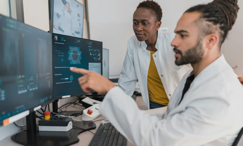
The group is a part of the department ‘imaging and pathology’ of the faculty of medicine, and focuses on medical physics aspects. Most of our research projects start from clinical remarks and/or complaints of the radiologists in the university hospital.
Topics of research include: quality evaluation of chest CT for lung cancer screening, optimisation of breast cancer screening techniques, patient dosimetry and automated dose and quality monitoring.
PhD students and master thesis students work under the guidance of Prof. Hilde Bosmans and Prof. Nicholas Marshall. Opportunities include the close cooperation with the radiology department, access to emerging x-ray equipment and patient images, a powerful software environment for online patient dose monitoring, and a framework of automatic image quality evaluation on CT scans.